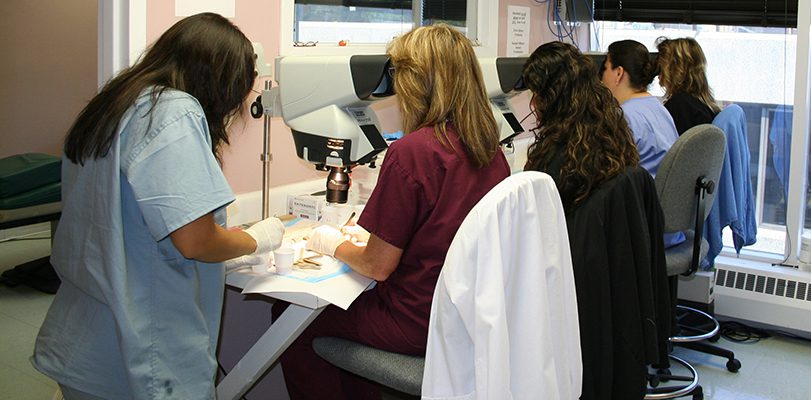
Why Use Microscopes For Hair Transplant Surgery?

For years, the use of microscopes in hair transplant surgery was an accepted standard. Using microscopes to review, inspect, and prepare grafts ensures the highest quality and best results. In recent years, however, an explosion of new clinics seeking an easy way to get into the hair restoration field have stopped using microscopes. These FUE-only clinics are unable to properly separate single-hair follicular unit grafts from multi-hair follicular unit grafts; these clinics are unable to inspect grafts to ensure the trauma from the FUE removal process is minimal and the grafts stand a good chance at surviving; and they are unable to refine grafts and remove excessive tissue before implantation. This leads to one inevitable conclusion: poorer results – particularly when multi-hair grafts are improperly identified as single-hair grafts and placed into the hairline where they appear bulky and create an artificial look.
Microscopes are required for FUT surgeries; the division of the strip down into the individual follicular units must be done under microscopic view. Thankfully, this means all grafts – from a strip – prepared by a skilled technician will be the highest quality by default. FUT with microscopic dissection places no trauma on the grafts, allows for perfect size trimming, removes all unwanted excess tissue, and allows the entire “pilo-sebaceous unit” (the follicular unit and its accompanying glands and attachments) to stay intact – ensuring better growth. FUE procedures, on the other hand, can technically be completed without microscopes. Since the follicular units are removed individually from the donor area and not as one unified piece, they do not need to be divided down – like the strip does in FUT. This means grafts can technically be removed via the FUE technique, examined with the naked eye or slight magnification, and implanted back into the recipient region. However, qualified clinics capable of performing both procedures and practiced in proper microscope do not perform FUE in this manner; instead, they review FUE grafts under microscopes before implanting in the recipient region. Clinics experienced in microscopic review are able to properly evaluate, classify, and prepare FUE grafts. This maximizes both the quality of the grafts and the chances of good growth.
Patients considering an FUE procedure must inquire whether or not microscopes are used during the procedure and what type of experience the clinic has with microscopic review. Ideally, the clinic would have ongoing experience with FUT graft preparation and possess the ability to translate this to FUE graft review and preparation.


